Understanding Tissue Dielectric Constant, Percentage Water Content and localized tissue-water assessment
LymphScanner gives clinicians a rapid, non-invasive way to assess local tissue water.
But what exactly does that mean?
Understanding the measurement does not require a deep knowledge of electromagnetic physics. The most important concept is simple:
Water strongly influences the electrical properties of tissue. LymphScanner uses this property to quantify water within a small, localized tissue volume.
This provides information that is fundamentally different from measuring limb circumference or total limb volume.
From TDC to tissue water
The technology behind LymphScanner is known as Tissue Dielectric Constant, or TDC.
A low-power electromagnetic signal is introduced into the tissue through the measurement probe. The interaction of that signal with the tissue depends strongly on the amount of water present.
The reflected signal is analyzed to determine the tissue's dielectric properties.
Because water has a much higher dielectric constant than most other tissue components, the measured TDC provides an index that is strongly dependent on the water contained within the measurement volume.
LymphScanner converts this information into an easy-to-interpret Percentage Water Content (PWC) value.
The clinician therefore does not need to interpret dielectric constants.
The clinical output is a localized tissue-water measurement.
What tissue is being measured?
This is an important distinction.
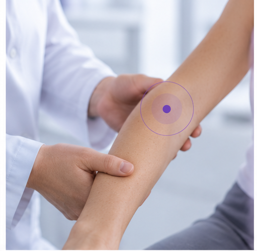
LymphScanner does not measure the entire arm or leg. It measures a small volume of tissue directly beneath the probe, primarily representing the skin and upper subcutaneous tissue.
LymphScanner has an effective measurement depth of approximately 2.5 mm.
This localization is one of the technology's most important clinical characteristics.
Consider two different questions:
“How much larger is this arm?”
and
“How much tissue water is present here?”
The first is a whole-limb or geometric question. The second is a local tissue question. LymphScanner is designed to help answer the second.
Free and bound water are included
At the approximately 300 MHz operating frequency used by the TDC measurement technology described in the research literature, the measurement is sensitive to both free and bound tissue water.
This means the measurement reflects the total water content within the measured tissue volume rather than only one narrowly defined fluid compartment.
For clinical education, the important message is:
LymphScanner measures localized total tissue water content within its measurement volume.
There is generally no need for the clinician to distinguish free and bound water during routine measurements.
Why can PWC differ between anatomical locations?
An isolated PWC value should always be interpreted in context.
Normal tissue-water values can vary between anatomical sites. Tissue composition, skin structure and the amount of subcutaneous tissue within the measurement volume all influence the absolute reading.
Research has repeatedly demonstrated anatomical-site differences in TDC values.
This is one reason why a PWC value measured on the hand should not automatically be interpreted in exactly the same way as the same numerical value measured on the forearm, breast or abdomen.
It is also one reason why matched anatomical comparisons and ratios can be particularly useful whenever an appropriate reference site is available.
Why ratios are powerful
Suppose the clinician measures corresponding sites on two arms.
Reference arm:
PWC = 32%
At-risk arm:
PWC = 40%
The ratio is:
40 / 32 = 1.25
Rather than asking whether 40% is intrinsically “high,” the ratio asks:
How different is the tissue water at this site compared with the corresponding reference tissue?
A ratio of 1.25 means that the measured local tissue-water value is 25% higher at the numerator site than at the reference site.
This does not mean that the entire arm contains 25% more fluid.
It describes a local difference at the measured sites.
Ratios help control normal physiological variation
Absolute tissue-water values can change for reasons unrelated to lymphedema.
One particularly useful TDC study followed measurements from morning to evening. Absolute TDC values changed during the day: upper-body measurements tended to decrease while calf values tended to increase.
In contrast, inter-side TDC ratios remained stable over the 13-hour observation period.
Other studies have found inter-arm ratios to be relatively robust to factors such as anatomical location, probe differences and handedness.
This provides an important practical reason for using ratios whenever a valid corresponding reference site is available:
The patient's own corresponding tissue becomes the reference.
This is valuable even when pre-operative baseline measurements are available.
Baseline values remain extremely useful for longitudinal surveillance, but a ratio adds simultaneous internal normalization at each visit.
What does research tell us about the inter-arm ratio?
The largest reference dataset currently available in our evidence base analyzed bilateral forearm TDC measurements from 403 healthy women.
Instead of defining the ratio according to dominant and non-dominant arms, the analysis calculated the higher TDC value divided by the lower TDC value.
Only 0.74% of the healthy women had an inter-arm ratio greater than 1.20.
This provides a useful way to understand the 1.20 reference level.
It is not a biological switch where lymphedema suddenly begins.
Rather:
A ratio around or above 1.20 represents a degree of local tissue-water asymmetry that is uncommon in healthy arms.
Clinical interpretation should still consider the patient's history, symptoms, physical examination and other relevant measurements.
In the United States, LymphScanner's cleared indication should also be followed precisely when the device is used clinically. The device supports local assessment of tissue-water differences between affected and contralateral non-affected arm tissues to aid clinical judgment of unilateral lymphedema in adult women; it is not intended to make a diagnosis or predict arm lymphedema.
Local measurement creates another opportunity: mapping
Because each measurement represents a localized area, measurements can be repeated across different anatomical regions.
Instead of assuming that tissue water is distributed uniformly, the clinician can investigate the tissue site by site.
This is particularly relevant because research using lymphofluoroscopy shows that lymphatic dysfunction and dermal backflow can have regional patterns. Local water-content measurements have shown meaningful agreement with dermal-backflow findings at several anatomical sites.
LymphScanner does not visualize lymphatic vessels or dermal backflow.
Instead, it provides another piece of information:
ICG lymphofluoroscopy → Where is lymphatic transport abnormal?
LymphScanner → Where is local tissue water increased?
Understanding this distinction is essential - and it also explains why localized tissue-water mapping is such an interesting area for clinical assessment.
Selected references
Mayrovitz HN. Assessing Free and Bound Water in Skin at 300 MHz Using Tissue Dielectric Constant Measurements with the MoistureMeterD. 2015.
Mayrovitz HN, Weingrad DN, Brlit F, Lopez LB, Desfor R. Tissue Dielectric Constant (TDC) as an Index of Localized Arm Skin Water: Differences Between Measuring Probes and Genders. Lymphology. 2015.
Mayrovitz HN, Weingrad DN, Lopez L. Tissue Dielectric Constant as an Index of Skin Water in Women With and Without Breast Cancer: Upper Limb Assessment via a Self-Contained Compact Measurement Device. Lymphology. 2016.
Mayrovitz HN. Diurnal Changes in Local Skin Water Assessed via Tissue Dielectric Constant at 300 MHz. Biomedical Physics & Engineering Express. 2017.
Mayrovitz HN. Assessing Unilateral Arm Lymphedema Status Using Interarm Tissue Dielectric Constant Ratios. Dataset of 403 healthy women.
Indication of Use
FDA clearance 510(k) in USA: LymphScanner is a device utilizing inter-aim ratios of tissue dielectric constant (TDC) that supports local assessment of tissue water differences between affected and contralateral non-affected arm tissues to aid in forming a clinical judgment of unilateral lymphedema in adult women. (K220557)
EU: LymphScanner is used to aid in forming a clinical judgement of edema at any soft tissue site of the body
Key takeaways
LymphScanner measures local tissue water content. It does not measure total limb volume.
The measurement is superficial and localized. LymphScanner assesses approximately the first 2.5 mm of skin and upper subcutaneous tissue.
PWC makes TDC clinically accessible. The underlying dielectric measurement is translated into Percentage Water Content (PWC).
Absolute PWC depends on location and physiology. A value should not be interpreted without considering where and how it was measured.
Ratios provide internal comparison. Comparing corresponding anatomical sites helps identify tissue-water asymmetry and reduces the influence of some normal physiological variation.
Multiple local measurements can reveal a pattern. This makes it possible to think beyond “the limb” and investigate specific areas of interest.

