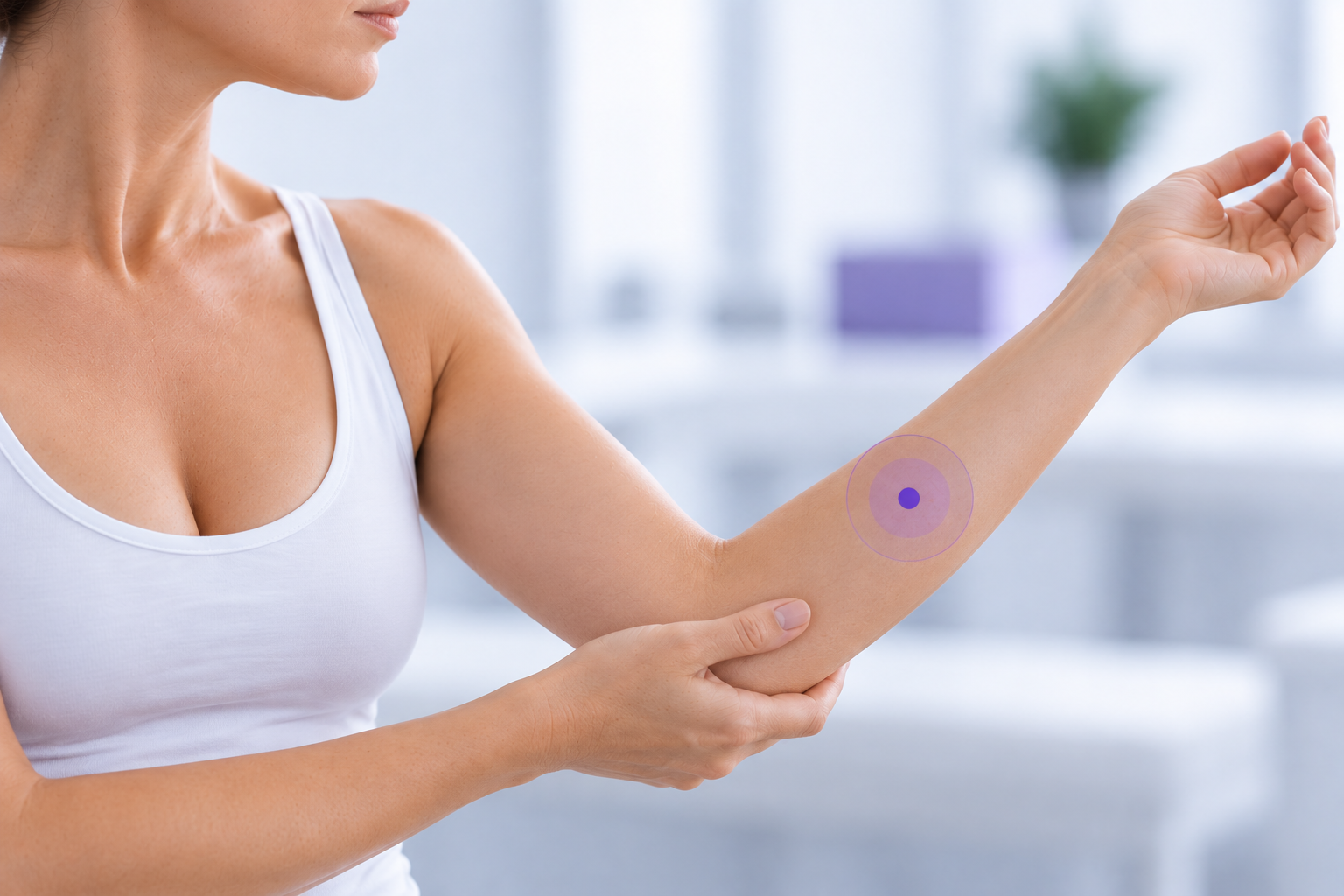
Yes. Fibrosis does not change the LymphScanner measurement principle—it changes the tissue being measured.
A common question about LymphScanner is:
“Does TDC still work when the tissue becomes fibrotic?”
The answer is yes.
LymphScanner uses Tissue Dielectric Constant (TDC) technology to measure local superficial tissue water content.
Fibrosis does not stop the TDC measurement principle from working. When appropriate probe-to-skin contact can be achieved at a suitable measurement site, LymphScanner continues to measure the dielectric properties of the superficial tissue beneath the probe.
However, as lymphedema progresses, the tissue itself changes.
This distinction is important because a limb can remain highly swollen - or become even larger - while fibrosis, skin thickening, adipose deposition and other structural changes increasingly affect the superficial tissue being measured.
As a result, local superficial tissue water content does not necessarily increase in parallel with overall limb swelling.
This can explain why LymphScanner values may be lower in advanced fibrotic tissue than during an earlier stage when superficial fluid accumulation was particularly prominent.
The measurement principle stays the same. The tissue does not.
What does LymphScanner actually measure?
LymphScanner measures local superficial tissue water content.
The measurement has an effective depth of approximately 2.5 mm and represents a small, localized superficial tissue volume beneath the probe.
Water strongly influences the dielectric properties of biological tissue. As the proportion of water within the measured tissue volume increases, the TDC value increases.
LymphScanner converts this dielectric information into Percentage Water Content, or PWC.
PWC therefore provides information about how much water is present within the local superficial tissue volume being measured.
It is not a direct measurement of:
- fibrosis
- tissue stiffness
- skin thickness
- limb volume
- lymphatic flow
This distinction becomes increasingly important as lymphedema progresses.
What happens in early lymphedema?
Early lymphatic dysfunction is strongly associated with changes in fluid transport.
When lymphatic outflow becomes insufficient, fluid can accumulate within the interstitial tissue. Altered superficial lymphatic transport can also produce dermal backflow.
Importantly, these changes can initially be localized and patchy rather than uniformly distributed throughout the limb.
Anatomical and ICG research describes dermal backflow beginning as small localized areas over compromised lymphatic vessels. Skin water content in dermal-backflow areas has been shown to be higher than in unaffected skin.
Johansson and Blom similarly describe early lymph-fluid accumulation as potentially non-uniform and identify TDC/PWC as a method for assessing local tissue water content in the skin and upper subcutis.
At this stage, the relationship is relatively intuitive:
Restricted lymphatic outflow
↓
Altered superficial lymphatic drainage / dermal backflow
↓
Localized superficial fluid accumulation
↓
Local superficial tissue water content increases
↓
PWC increases
Substantial structural tissue remodeling may not yet be present.
This helps explain why localized TDC measurement can be particularly informative in early and mild disease.
What happens as swelling becomes more persistent?
As lymphatic dysfunction persists, swelling can continue to develop.
The amount of excess fluid within the affected limb or body region may continue to increase. The limb may become visibly larger and the swelling more persistent.
At the same time, however, another process begins to become increasingly important:
the tissue itself starts to remodel.
Persistent lymphatic congestion is associated with protein-rich interstitial fluid stasis and chronic inflammation.
Over time, this environment can contribute to changes such as increased extracellular matrix deposition, fibrosis, adipose accumulation, skin thickening and other structural alterations.
The affected limb can therefore continue to increase in size while its tissue composition is simultaneously changing.
This gives us two processes that can occur at the same time:
Overall swelling can remain high or continue to increase.
while
The composition of the superficial tissue measured by LymphScanner can progressively change.
That distinction is central to understanding TDC measurements in fibrotic lymphedema.
Whole-limb swelling and superficial tissue water content are not the same thing
Imagine a patient whose arm becomes progressively larger.
The increase in limb size may reflect a combination of:
excess fluid + fibrosis + connective-tissue remodeling + adipose deposition + skin changes
LymphScanner does not measure all of these components or the total amount of fluid within the entire limb.
It asks a much more localized question:
“What is the water content of the superficial tissue at this specific measurement site?”
This means that a very large or severely swollen limb does not necessarily have extremely high PWC at every superficial measurement location.
Likewise, if PWC at a particular site decreases as disease progresses, it does not necessarily mean that the total swelling of the limb has decreased.
Overall swelling may continue to progress while local superficial tissue water content follows a different trajectory as the superficial tissue remodels.
Why might PWC decrease as fibrosis and tissue remodeling progress?
This is an important question - and one where physiological interpretation needs to be distinguished from direct clinical evidence.
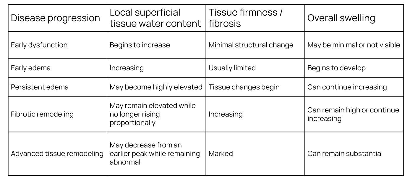
Localized tissue-water content and tissue-firmness changes during lymphedema progression, LymphScanner values are expected to initially rise as superficial tissue fluid increases.
As swelling becomes more persistent, superficial tissue-water values may remain high while tissue firmness and structural tissue changes begin to increase.
With advanced tissue remodeling and fibrosis, PWC may decrease from its earlier peak while tissue firmness continues to increase.
Several tissue changes provide plausible explanations for this pattern.
1. The superficial measurement volume changes in composition
Early in the disease process, increased fluid can represent a prominent change within the superficial tissue being measured.
As disease progresses, the same superficial tissue compartment may increasingly contain structural changes associated with chronic lymphedema.
Fibrosis involves increased deposition and remodeling of extracellular matrix. Skin and subcutaneous tissue can thicken and become structurally altered, while adipose tissue may also contribute increasingly to the affected tissue.
The approximately 2.5 mm superficial tissue volume measured later in the disease may therefore be very different from the tissue measured earlier.
This is the essential principle:
Fibrosis does not change how LymphScanner measures - it changes the composition and structure of the tissue being measured.
2. Superficial water may represent a different proportion of the remodeled tissue
As collagen-rich extracellular matrix and other structural components become more prominent, water may represent a different proportion of the local superficial measurement volume.
This does not mean that the entire limb has become less fluid-rich.
There may still be substantial excess fluid elsewhere within the limb or at deeper tissue levels.
Instead, it means that the local superficial tissue sampled by LymphScanner may no longer contain the same proportion or distribution of water that it did when superficial fluid accumulation was more prominent.
This is an important difference.
A lower PWC can coexist with persistent or increasing overall swelling.
3. Fibrotic remodeling may change the space available for superficial interstitial fluid
Progressive tissue remodeling may reduce the open space available for freely accumulating interstitial fluid within the superficial measurement volume.
In earlier stages, increasing local superficial fluid accumulation is expected to increase PWC.
As structural remodeling progresses, however, the local relationship between tissue structure and water changes.
PWC may therefore remain abnormal but no longer continue rising with disease progression. In advanced tissue, it may decrease from an earlier maximum even while the limb remains substantially swollen.
Again, this describes the superficial tissue measurement.
It should not be interpreted as evidence that the entire limb contains less excess fluid.
4. Skin thickening changes the superficial tissue being sampled
Advanced lymphedema can produce marked changes in the skin itself.
The epidermis and dermis may become thicker and structurally altered, while fibrosis and other chronic tissue changes increasingly affect the superficial compartment.
This is particularly relevant to LymphScanner because its measurement is intentionally superficial.
The device continues to interrogate approximately the same measurement depth, but the anatomy and composition contained within that depth can change as the disease progresses.
This provides another reason why PWC should not be expected to simply follow whole-limb size.
5. Fluid distribution can remain highly heterogeneous
Advanced lymphedema is not necessarily uniform.
One region may have substantial superficial fluid accumulation, while another may have more pronounced fibrotic or adipose remodeling.
Similarly, fluid may be distributed differently between superficial and deeper tissue compartments.
A single local PWC measurement therefore describes that measurement site only, not the total fluid burden of the limb.
This is another reason localized measurements should be interpreted according to anatomical location and, where appropriate, followed at consistent sites over time.
Does dermal backflow disappear when fibrosis develops?
Not necessarily.
In early lymphedema, dermal backflow can begin as small localized areas over compromised lymphatic vessels.
As disease progresses, dermal-backflow areas can become more extensive. In advanced lymphedema, superficial collecting vessels may become increasingly damaged while dilated lymphatic capillaries and pre-collectors contribute to alternative lymph transport.
So we should not explain decreasing PWC by saying that dermal backflow simply disappears.
Instead:
The relationship between dermal backflow, superficial local tissue water content and tissue structure becomes more complex as lymphedema progresses.
This is also why evidence relating dermal backflow to elevated local tissue water content should not be interpreted as meaning every dermal-backflow region will have the same PWC at every stage of disease.
Does TDC still work in fibrotic tissue?
Yes.
Fibrosis does not switch off the physical measurement principle of TDC.
When the probe can be appropriately positioned with good skin contact at a suitable measurement site, LymphScanner continues to assess local superficial tissue water content.
What changes is the tissue being measured.
This is why an advanced fibrotic limb can present an initially surprising combination:
The limb may be very large.
Overall swelling may still be substantial.
Fibrosis and tissue remodeling may be advanced.
Yet superficial PWC at a particular location may be lower than it was earlier in the disease process.
These observations are not necessarily contradictory.
They describe different characteristics and compartments of the affected tissue.
A lower PWC in advanced fibrotic tissue does not mean that LymphScanner has stopped measuring correctly. It may reflect a real change in the water content and composition of the superficial tissue being measured.
A useful way to understand the relationship
Localized superficial tissue water content, tissue firmness and overall swelling should not necessarily be expected to follow the same trajectory as lymphedema progresses.
The key concept is not that advanced lymphedema contains less fluid overall.
It is that:
Superficial tissue water content and total limb swelling can follow different trajectories as tissue remodeling progresses.
What if the limb is very swollen but PWC is only moderately elevated?
This can be physiologically understandable.
Whole-limb size reflects everything contributing to the volume of the limb.
LymphScanner measures something much more specific: local superficial tissue water content within approximately 2.5 mm of the measurement site.
A large limb may contain substantial overall fluid while also containing increased fibrotic tissue, adipose tissue, thickened skin and other structural changes.
Some of that fluid may also be distributed outside the superficial tissue volume sampled at a particular LymphScanner measurement site.
Therefore:
A large limb does not require a high superficial PWC at every location.
And equally:
A moderate PWC does not mean there is only moderate lymphedema overall.
PWC should be interpreted as what it is: a localized measurement of superficial tissue water content.
Fibrotic tissue can still have elevated PWC
Advanced fibrotic tissue may continue to show elevated superficial tissue-water values compared with unaffected tissue, even when PWC has decreased from an earlier peak.
So fibrosis and elevated PWC can coexist.
A region can be:
highly fibrotic
and
still have elevated local superficial tissue water content.
These findings describe different properties of the same tissue.
PWC and tissue firmness answer different questions
This leads to an important clinical distinction.
LymphScanner asks:
How much water is present in this local superficial tissue volume?
A tissue-firmness assessment asks:
How resistant is this tissue to mechanical deformation?
The two properties can change differently over time.
This is why:
High PWC does not prove fibrosis.
Lower PWC does not exclude fibrosis.
Increasing tissue firmness does not mean TDC has become unreliable.
And:
A very swollen limb does not require an extremely high PWC at every superficial location.
What does this mean when monitoring treatment?
The same principle matters during longitudinal follow-up.
Treatment may influence different tissue properties differently.
A clinician may observe changes in:
- local superficial tissue water content
- tissue firmness
- tissue mobility
- limb volume
- fluid distribution
- symptoms
These do not necessarily change simultaneously or in the same direction.
If PWC decreases, the most direct interpretation is:
Local superficial tissue water content has decreased at this measurement site.
It does not automatically mean:
“Fibrosis has decreased.”
Likewise, if tissue becomes softer while PWC remains elevated, the measurements are not necessarily conflicting.
They are measuring different properties.
This is why longitudinal measurements are most informative when the same anatomical locations are followed consistently and interpreted together with the clinical picture.
How should clinicians interpret TDC in fibrotic lymphedema?
The simplest approach is to stay close to what each measurement actually tells us.
Measure water as water.
LymphScanner provides information about local superficial tissue water content.
Assess fibrosis as fibrosis.
Tissue firmness, pitting, skin characteristics and other structural changes require their own clinical or objective assessment.
Assess swelling as swelling.
Limb volume and circumference provide information about the overall dimensions of the affected region.
These measurements can complement one another precisely because they do not measure the same thing.
Selected references
Suami H. Anatomy and Physiology of the Lymphatic System and Structural Changes in Lymphedema. 2020.
Johansson KI, Blom KY. Prevention of Breast Cancer–Related Arm Lymphedema. 2025.
Thomis S, et al. Clinical assessment of patients with breast cancer-related lymphedema: comparison of methods with lymphofluoroscopy.
Mayrovitz HN, Yzer JA. Local Skin Cooling as an Aid to the Management of Patients with Breast Cancer Related Lymphedema and Fibrosis of the Arm or Breast. Lymphology. 2017.
Mayrovitz HN, Shams E, Astudillo A, et al. Tissue Dielectric Constant and Skin Stiffness Relationships in Lower Extremity Lymphedema.
Yu Z, et al. Assessment of Skin Properties in Chronic Lymphedema: Measurement of Skin Stiffness, Percentage Water Content, and Transepidermal Water Loss.
Indication of Use
FDA clearance 510(k) in USA: LymphScanner is a device utilizing inter-aim ratios of tissue dielectric constant (TDC) that supports local assessment of tissue water differences between affected and contralateral non-affected arm tissues to aid in forming a clinical judgment of unilateral lymphedema in adult women. (K220557)
EU: LymphScanner is used to aid in forming a clinical judgement of edema at any soft tissue site of the body
Key takeaways
Yes. TDC measurement works in fibrotic tissue
- Fibrosis does not change the LymphScanner measurement principle. It changes the tissue being measured.
LymphScanner measures local superficial tissue water content.
- It does not measure total limb fluid, limb volume or fibrosis.
Early lymphatic dysfunction can produce prominent superficial tissue-water changes.
- Localized fluid accumulation and dermal backflow can increase superficial tissue water content before substantial structural remodeling has developed.
Overall swelling can continue to increase while the superficial tissue changes.
- Persistent swelling and progressive fibrosis, skin thickening, adipose deposition and other tissue remodeling can occur at the same time.
Superficial PWC and whole-limb swelling do not have to follow the same trajectory.
- PWC may stop increasing or decrease from an earlier peak even while overall swelling remains substantial or continues to progress.
This does not mean LymphScanner has stopped working.
- It may reflect a real change in the composition and water content of the superficial tissue being measured.
Advanced lymphedema should not simply be described as “less fluid-dominant.”
- The limb can contain substantial excess fluid. The important distinction is that the superficial tissue within the LymphScanner measurement volume may have changed considerably.
Fibrotic tissue can still have elevated PWC.
- Fibrosis and elevated superficial tissue water content can coexist.
PWC is not a fibrosis score or a measure of total disease severity.
- It should be interpreted only as an objective measurement of local superficial tissue water content.
Fibrosis does not change how LymphScanner measures - it changes the tissue being measured.
Overall swelling may continue to progress, while local superficial tissue water content follows a different trajectory as the tissue remodels.

