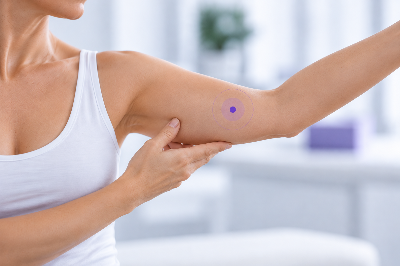
Understanding why local tissue changes matter in lymphedema assessment
For many years, lymphedema assessment has focused heavily on a simple question:
Has the limb become larger?
Circumference measurements, limb volume and water displacement remain valuable clinical tools. But understanding of lymphedema has evolved.
Lymphedema does not necessarily begin as uniform enlargement of an entire limb. Particularly in earlier or milder disease, lymphatic dysfunction and fluid accumulation can be regional and heterogeneous.
This changes the question clinicians may want to ask.
Instead of only:
“Is the limb bigger?”
we can also ask:
“Where is tissue water changing?”
That distinction is important for understanding the role of localized tissue-water assessment.
Lymphedema begins with impaired lymphatic transport
The lymphatic system helps maintain tissue-fluid balance by transporting fluid and macromolecules away from the interstitial space.
Cancer treatment can disrupt this system. Surgery may remove or damage lymphatic structures, while radiation can affect lymphatic transport and surrounding tissues.
When lymphatic transport capacity is insufficient for the lymphatic load, fluid can accumulate in the tissues.
In breast cancer-related lymphedema, this may occur in the arm, hand, breast or trunk, and the location and extent of fluid accumulation can vary considerably between patients.
This is one reason why assessing lymphedema solely through whole-limb size can provide only part of the clinical picture.
Early changes may be localized
One of the most important developments in understanding of lymphedema comes from lymphatic imaging.
Near-infrared fluorescence imaging, often referred to as ICG lymphography or lymphofluoroscopy, can visualize superficial lymphatic transport.
Healthy lymphatic transport typically appears as relatively linear lymphatic pathways.
When lymphatic transport becomes impaired, characteristic abnormal patterns may develop. These are commonly described as splash, stardust and diffuse patterns, representing progressively altered superficial lymphatic transport and dermal backflow.
Importantly, these changes are not necessarily distributed uniformly throughout the limb.
Research summarized by Johansson and Blom describes how fluid accumulation in the initial stages of lymphedema can be localized within the subcutaneous tissues rather than evenly distributed throughout the arm.
That has an important practical consequence:
A patient can have clinically relevant local tissue changes even when the entire limb does not yet appear substantially larger.
The skin and subcutaneous tissue matter
Lymphedema is therefore not simply a question of total limb volume.
The disease involves changes within the tissue itself.
Early stages are characterized primarily by fluid accumulation. As lymphedema progresses, chronic inflammation and tissue remodeling can contribute to changes such as increased skin and subcutaneous thickness, adipose deposition and fibrosis.
This is why examination of the skin and subcutaneous tissues has an important role alongside limb-volume assessment.
Johansson and Blom recommend that diagnosis should not depend solely on volume differences. Clinical examination of skin and subcutaneous thickness, patient symptoms and methods assessing tissue fluid can provide complementary information.
Different methods answer different clinical questions

These methods should not necessarily be expected to produce identical findings because they measure different aspects of the condition.
They can be complementary.
Dermal backflow provides an important clue
A particularly interesting connection has been demonstrated between lymphatic imaging and local tissue-water assessment.
In a study of 45 patients with stage I–IIb breast cancer-related lymphedema, Thomis and colleagues compared clinical measurements at specific anatomical locations with dermal backflow visualized using lymphofluoroscopy.
Local water content showed an overall association with the presence of dermal backflow. At several regions - including the hand, ventral and dorsal forearm, and ventral upper arm - water-content findings correlated with the lymphofluoroscopic findings.
The authors concluded that skinfold thickness, water content and lymphedema volume were among the most appropriate clinical measurements for detecting dermal backflow.
This does not mean that tissue-water measurement images lymphatic vessels or directly detects dermal backflow.
ICG lymphofluoroscopy and TDC measure different things.
ICG visualizes lymphatic transport patterns.
TDC measures local tissue water.
But the relationship between disturbed lymphatic transport and increased local tissue water helps explain why localized tissue-water measurement can provide useful information in lymphedema assessment.
Regions of dermal backflow (green areas) illustrate lympedema in skin and LymphScannercan quantitate tissue water locally at each site
From measuring a limb to understanding a tissue pattern
This leads to a different way of thinking about lymphedema assessment.
A whole-limb measurement gives us a global view.
A localized measurement allows us to investigate individual areas of interest.
For example, instead of taking one measurement and assuming it represents the entire arm, a clinician can assess multiple anatomical regions:
Hand → forearm → elbow region → upper arm → axillary region
The purpose is not simply to collect more numbers.
It is to understand whether tissue-water abnormalities are localized, widespread or changing over time.
The same principle becomes particularly relevant outside the limbs—in areas such as the breast and trunk, where conventional circumference or limb-volume measurements are inherently difficult or impossible to apply.
Why this matters for early assessment
Early or mild lymphedema may not produce obvious symptoms.
Johansson and Blom note that patients with early mild breast cancer-related arm lymphedema do not necessarily experience subjective arm symptoms and may therefore be unaware of its onset. They recommend surveillance and the combined use of local tissue-water assessment, volume measurement and clinical examination when evaluating early mild disease.
Their review also describes evidence that TDC identified lymphedema at a lower relative limb volume and earlier after surgery than water-displacement measurement alone in one study.
The important concept is therefore not simply measuring earlier in time.
It is trying to recognize abnormal change earlier in the biological progression of the condition.
The clinical mindset shift
Traditional question
Is the limb bigger?
Additional question enabled by localized tissue-water assessment
Is tissue water abnormal somewhere within the area at risk?
Both questions can matter.
But they are not the same question.
Selected references
Thomis S, Dams L, Fourneau I, et al. Correlation Between Clinical Assessment and Lymphofluoroscopy in Patients with Breast Cancer-Related Lymphedema: A Study of Concurrent Validity. Lymphatic Research and Biology. 2020.
Johansson KI, Blom KY. Prevention of Breast Cancer–Related Arm Lymphedema. 2025.
Mayrovitz HN. Measuring Breast Cancer-Related Lymphedema. 2022. [This is among the uploaded evidence base.]
Toro C, Markarian B, Mayrovitz HN. Breast Cancer-Related Lymphedema Assessed via Tissue Dielectric Constant Measurements. Cureus. 2024.
Indication of Use
FDA clearance 510(k) in USA: LymphScanner is a device utilizing inter-aim ratios of tissue dielectric constant (TDC) that supports local assessment of tissue water differences between affected and contralateral non-affected arm tissues to aid in forming a clinical judgment of unilateral lymphedema in adult women. (K220557)
EU: LymphScanner is used to aid in forming a clinical judgement of edema at any soft tissue site of the body
Key takeaways
Lymphedema can be regional. Tissue-fluid accumulation does not necessarily occur uniformly throughout a limb.
Early disease may be difficult to see. Local tissue changes may exist before substantial whole-limb changes becomes apparent.
Skin and subcutaneous tissue are important. Lymphedema involves changes within these tissues, not simply external limb geometry.
Different measurement methods provide different information. Local tissue-water assessment complements rather than replaces volume measurements and clinical examination.
Think in areas of interest. Understanding where tissue water is changing can add a new dimension to surveillance, clinical assessment and treatment monitoring.

