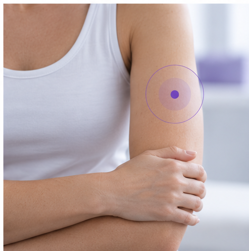
Early lymphedema is not always a whole-limb problem. Changes in superficial lymphatic transport can be localized, patchy and dynamic—helping explain why local superficial tissue water content can provide information that whole-limb measurements may not fully capture.
When people think about lymphedema, they often imagine a limb gradually becoming uniformly swollen.
The reality is more complex.
The superficial lymphatic system is organized into distinct pathways and territories. After cancer treatment, some pathways may remain functional, some may become restricted, and the lymphatic system may adapt through alternative or compensatory drainage routes.
As a result, early or mild lymphedema may appear as a regional tissue problem before it becomes an obvious whole-limb problem. These regional changes can include localized increases in superficial tissue water content associated with areas of altered lymphatic drainage.
How does normal superficial lymphatic drainage work?
The superficial lymphatic system begins close to the skin surface.
Lymphatic capillaries are located within the skin. These connect to pre-collectors, which in turn drain into larger superficial lymph collecting vessels in the subcutaneous tissue.
The collecting vessels transport lymph toward regional lymph nodes and eventually back into the venous circulation.
Unlike the blood circulatory system, lymphatic transport does not rely on a central pump. Flow is supported by the intrinsic contraction of lymphatic collecting vessels, one-way valves, muscle movement, respiration and other external forces.
The superficial lymphatic system is also organized into anatomical territories. Groups of superficial lymphatic vessels drain defined skin regions toward particular first-tier lymph nodes. This organization forms the basis of the lymphosome concept.
This matters because the superficial lymphatic system should not be thought of as one uniform drainage compartment.
What changes after cancer treatment?
Cancer treatment can alter lymphatic transport.
Lymph node surgery and radiotherapy can restrict or interrupt existing drainage pathways. But this does not necessarily mean that all lymphatic drainage simply stops.
The original drainage route may remain partly functional, while collateral or alternative drainage pathways may develop. When flow through superficial collecting vessels becomes restricted, dermal backflow can also create connections between compromised and still-functioning lymphatic pathways.
This is why describing lymphedema simply as a “blocked lymphatic system” can be misleading.
For example, anatomical and ICG imaging research indicates that the ipsilateral axilla can continue to function as an important drainage pathway even after axillary lymph node dissection. The authors therefore suggest describing the pathophysiology as insufficient lymphatic drainage caused by restriction of outflow, rather than complete blockage of lymphatic drainage.
A useful way to think about this is:
Lymphedema develops when lymphatic outflow becomes insufficient for the fluid load, despite the body's attempts to maintain or reroute drainage.
The balance between impaired and compensatory pathways can differ between patients-and between regions within the same patient.
What is dermal backflow?
Dermal backflow is an important sign of altered superficial lymphatic transport.
When upstream flow through lymphatic collecting vessels becomes restricted, lymph can reflux from the collecting vessels into the superficial lymphatic network within the skin.
This is associated with dilation of lymphatic capillaries and pre-collectors and is known as dermal backflow. It can be directly visualized using indocyanine green, or ICG, lymphography.
Importantly, dermal backflow does not necessarily involve the entire limb.
In early lymphedema, it can begin as a small, localized area over compromised lymphatic vessels. Nearby lymphatic pathways may still remain patent and functional.
As lymphatic dysfunction progresses and more collecting vessels become affected, the area of dermal backflow can become larger and more widespread.
This helps explain one of the most important features of early disease:
Early lymphatic dysfunction can be highly regional.
Dermal backflow reflects dysfunction - but can also support compensatory drainage
Dermal backflow is often described primarily as a sign of lymphatic dysfunction.
The anatomy shows a more dynamic picture.
In mild to moderate lymphedema, dermal backflow may form a bridge between compromised and still-patent lymphatic vessels, allowing lymph to reach functioning drainage pathways.
As superficial collecting vessels deteriorate further, the dermal lymphatic network may increasingly serve as an alternative route for lymph transport.
This reinforces an important concept:
The lymphatic system in lymphedema is not simply blocked. It is impaired, but it may also be adapting, compensating and rerouting lymph.
This is one reason why lymphatic dysfunction can present differently from one patient to another - and from one region to another.
Why does dermal backflow matter for tissue water content?
Dermal backflow changes not only the pattern of lymphatic transport but also the tissue through which that altered transport occurs.
When upstream lymphatic outflow becomes restricted, lymph can reflux into the superficial lymphatic network within the skin. Anatomical and imaging research shows that skin water content is higher in dermal-backflow areas than in unaffected tissue.
This creates an important connection between lymphatic dysfunction and localized tissue-water measurement:
Restricted lymphatic outflow
↓
Dermal backflow / altered superficial lymphatic drainage
↓
Localized increase in superficial tissue water content
↓
Tissue-water abnormality detectable with TDC
This connection is particularly important for understanding what LymphScanner measures.
What does research show about local tissue water content and dermal backflow?
The relationship between dermal backflow and local tissue water content has also been investigated clinically.
Thomis and colleagues compared lymphofluoroscopy findings with different clinical assessment methods in patients with breast cancer-related lymphedema.
Local tissue-water content measurements showed an association with dermal backflow at several anatomical regions, including the hand, forearm and upper arm.
This does not mean that tissue-water measurement and lymphatic imaging measure the same thing.
They do not.
ICG lymphography visualizes lymphatic transport and dermal backflow.
TDC measures local superficial tissue water content.
The relationship between the two is clinically important because altered superficial lymphatic transport can create localized increases in tissue water content that can be detected and quantified with TDC.
How can LymphScanner help identify dermal-backflow areas?
LymphScanner does not measure dermal backflow itself.
Dermal backflow describes an alteration in lymphatic transport. ICG lymphography can directly visualize this phenomenon and provide information about lymphatic pathways and the movement of lymph.
LymphScanner measures something different: local tissue water content.
This distinction is important because the two phenomena are related.
Anatomical and imaging studies have shown that tissue water content is higher in areas of dermal backflow than in unaffected tissue. Clinical research has also demonstrated an association between local tissue-water measurements and dermal-backflow patterns identified with lymphatic imaging.
LymphScanner can therefore help identify and localize areas of elevated superficial tissue water content that may correspond to dermal-backflow areas.
In practical terms:
ICG visualizes dermal backflow and lymphatic drainage patterns.
LymphScanner detects and quantifies the localized superficial tissue-water content abnormalities that can be associated with altered lymphatic drainage and dermal backflow.
LymphScanner does not show how lymph is flowing, where it is being rerouted or which individual lymphatic vessels remain patent.
Instead, it provides information about the local tissue-water consequence of altered lymphatic transport.
An area with elevated local PWC may therefore represent tissue affected by dermal backflow or other lymphatic dysfunction. The measurement should be interpreted as evidence of a localized tissue-water abnormality, rather than direct visualization or confirmation of dermal backflow.
Why superficial tissue-water measurement matters
The superficial measurement is important because of where many of the relevant lymphatic structures are located.
Lymphatic capillaries and pre-collectors are located within the skin, while superficial lymph collecting vessels run within the subcutaneous tissue. In lymphedema, dermal backflow involves reflux into this superficial lymphatic network.
LymphScanner has an effective measurement depth of approximately 2.5 mm, providing a localized measurement of superficial tissue water content.
This places the LymphScanner measurement within the superficial tissue compartment where dermal lymphatic abnormalities and associated tissue-water changes can occur.
Importantly, LymphScanner does not measure the lymphatic vessels themselves.
It measures local superficial tissue water content within a tissue compartment that is highly relevant to lymphedema pathophysiology.
Early detection means finding change while the burden is still low
The regional nature of early lymphatic dysfunction is particularly important for prospective surveillance.
Early detection does not only mean measuring soon after surgery.
It also means recognizing abnormal change early in the disease process, while the overall tissue and symptom burden may still be low.
Prospective ICG research following patients after breast cancer surgery has demonstrated that lymphatic dysfunction can be identified before significant limb-volume changes are present.
This helps explain why relying only on whole-limb enlargement can miss an important part of early disease.
A limb does not necessarily need to become globally larger before lymphatic dysfunction begins affecting the tissue.
The earlier question may therefore be not only “Is the limb getting bigger?” but also “Is local superficial tissue water content becoming abnormal somewhere within the tissue at risk?”
That is where localized measurement can add information.
The breast provides another example of regional adaptation
Breast lymphedema also demonstrates how regional lymphatic drainage can change after treatment.
ICG studies in women with breast lymphedema have identified dermal backflow together with collateral and alternative drainage pathways.
Instead of relying exclusively on the normal dominant pathway toward the ipsilateral axilla, drainage may involve parasternal, contralateral axillary, intercostal or clavicular regions.
Research in this area has also used localized TDC measurements to assess breast tissue water content.
The important clinical lesson is not that one specific alternative route always develops.
It is that:
Lymphatic adaptation is individual, regional and dynamic.
Why one measurement point may not be enough
Anatomical research provides another important insight.
Individual superficial lymphatic collecting vessels can serve relatively narrow skin territories. Comprehensive lymphatic imaging therefore requires assessment from multiple anatomical regions because evaluating only one or two areas may miss affected lymphatic pathways elsewhere.
LymphScanner does not identify individual lymphatic territories or collecting vessels.
However, the regional organization of the superficial lymphatic system helps explain why superficial tissue-water abnormalities may also be spatially heterogeneous.
A local tissue-water content measurement at one location should therefore not automatically be assumed to represent the entire limb or anatomical region.
This is why assessing multiple relevant areas of interest may provide a more complete picture than relying on a single measurement point.
From measuring limbs to identifying areas of interest
This evidence supports a broader clinical mindset.
Traditional whole-limb measurements often begin with the question:
“Is the limb larger?”
Localized tissue-water assessment allows another question:
“Where is superficial tissue water content abnormally elevated?”
Relevant areas of interest may be influenced by:
- the location of surgery
- lymph node procedures
- radiation fields
- patient-reported heaviness, fullness or tightness
- visible or palpable tissue changes
- previously identified abnormal measurement sites
Localized tissue-water measurements can then help clinicians investigate these areas systematically.
Identifying an area with elevated PWC does not reveal the underlying lymphatic pathway. However, it may help identify a region affected by altered lymphatic drainage, including a possible dermal-backflow area.
The goal is therefore not simply to find the highest PWC value.
The goal is to understand:
where local superficial tissue water content is elevated,
how it compares with appropriate reference tissue,
and how the pattern changes over time.
A practical clinical framework
Localized assessment can be thought of as:
SPOT
Identify areas with elevated local superficial tissue water content.
MAP
Assess multiple relevant areas to understand the regional distribution of tissue-water abnormalities.
COMPARE
Use an anatomically appropriate reference site whenever possible.
TRACK
Document important areas of interest and follow their tissue water content over time.
This framework is a clinical education concept based on the evidence for regional lymphatic dysfunction and localized tissue-water abnormalities. It is not intended to replace clinical examination or lymphatic imaging.
Selected references
Suami H, Shinaoka A. Anatomy and Structural Physiology of the Lymphatic System.
Suami H, Kato S. Anatomy of the Lymphatic System and Its Structural Disorders in Lymphoedema. Springer; 2018.
Suami H, Koelmeyer L, Mackie H, Boyages J. Patterns of lymphatic drainage after axillary node dissection impact arm lymphoedema severity: A review of animal and clinical imaging studies. Surgical Oncology. 2018;27:743–750.
Akita S, et al. Early Detection of Lymphatic Disorder and Treatment for Lymphedema following Breast Cancer. Plastic and Reconstructive Surgery. 2016.
Thomis S, et al. Clinical assessment of patients with breast cancer-related lymphedema: comparison of methods with lymphofluoroscopy.
Heydon-White A, Suami H, Boyages J, Koelmeyer L, Peebles KC. Assessing breast lymphoedema following breast cancer treatment using indocyanine green lymphography. Breast Cancer Research and Treatment. 2020.
Koelmeyer LA, et al. Personalizing Conservative Lymphedema Management Using Indocyanine Green-Guided Manual Lymphatic Drainage. Lymphatic Research and Biology. 2021.
Weber E, et al. Lymphatic Collecting Vessels in Health and Disease: A Review of Histopathological Modifications in Lymphedema. Lymphatic Research and Biology. 2022.
Indication of Use
FDA clearance 510(k) in USA: LymphScanner is a device utilizing inter-aim ratios of tissue dielectric constant (TDC) that supports local assessment of tissue water differences between affected and contralateral non-affected arm tissues to aid in forming a clinical judgment of unilateral lymphedema in adult women. (K220557)
EU: LymphScanner is used to aid in forming a clinical judgement of edema at any soft tissue site of the body
Key takeaways
Early lymphatic dysfunction can be regional.
Dermal backflow may begin as a small, localized abnormality while nearby lymphatic pathways remain functional.
Lymphedema is more than a blocked lymphatic system.
Restricted outflow can coexist with original, collateral and compensatory drainage.
Dermal backflow reflects altered lymphatic transport.
It develops when restricted upstream flow causes lymph to reflux into the superficial lymphatic network and may also contribute to compensatory drainage.
Dermal-backflow areas can contain elevated tissue water content.
Anatomical and imaging research shows higher skin water content in dermal-backflow areas than in unaffected tissue.
LymphScanner can help identify these localized tissue-water abnormalities.
Elevated local superficial tissue water content may correspond to an area affected by dermal backflow or other lymphatic dysfunction.
LymphScanner does not measure dermal backflow itself.
LymphScanner detects and quantifies the localized tissue-water content abnormalities that can be associated with altered lymphatic drainage and dermal backflow. But it does not visualize lymphatic vessels, lymph flow patterns or drainage direction.
Localized assessment complements whole-limb measurement.
A patient may have meaningful regional tissue-water abnormalities before substantial whole-limb swelling becomes apparent.
Early lymphedema may begin as a local tissue problem before it becomes a whole-limb problem.

